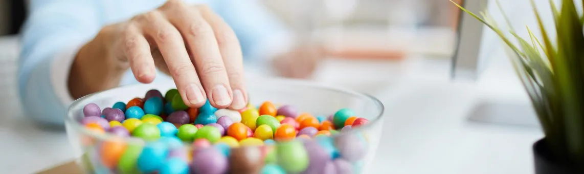
3D Printing

3D Printing is Making Inroads in Everyday Medicine.

3D printing — the revolutionary technology that can crank out a rocket nozzle the way a desktop printer can spit out a tax return — has captivated the imagination of engineers and researchers for more than a decade. In medicine, the technology is proving itself invaluable every day in a wide variety of surgical, research, and training applications.

What makes 3D printing so promising for medicine is not just its ability to build an object from scratch; it’s the way the technology can turn data from, say, a CT scan, into an exact match of a unique piece of a patient’s anatomy from virtually anywhere in the body. At UPMC, medical and bioengineering experts are finding ways to make this once exotic technology a cost-effective method for mapping tumors, performing reconstructive surgery, guiding ACL operations, and much more.

Never Miss a Beat!

Get Healthy Tips Sent to Your Phone!

Message and data rates may apply. Text the word STOP to opt out and HELP for help. Click here to view the privacy and terms.

Navigating a Kidney Tumor

One strong advocate of the use of 3D models in cancer surgery is Stephen V. Jackman, MD, a professor at the University of Pittsburgh School of Medicine and director of UPMC’s urology residency program. “CT scans and MRI images are limited,” says Dr. Jackman. “One of the best things a 3D model does is support patient education. A patient can hold it in his or her hand and understand what the operation is going to be like.”

Surgeons also benefit from the use of 3D models, he adds. “Unlike a CT image, a surgeon can orient the model exactly the way the kidney displays on the screen. This does a better job of helping us find a tumor, understand how deep it goes, remove it, and repair the kidney afterwards,” says Dr. Jackman.

A Dry Run For Delicate Reconstruction

In surgery to correct aural atresia, a rare condition in which a child is born without an ear canal, a 3D model serves as an accurate, realistic stand-in for the patient’s anatomy so that a surgeon can practice the delicate procedure before live surgery.

Noel Jabbour, MD, MS, director of the Congenital Ear Center at UPMC Children’s Hospital of Pittsburgh, finds 3D printing “game-changing” for this operation, which requires precise bone removal in a complex area crowded with facial nerves, jaw anatomy, and delicate inner ear structures. “Until now there’s been no way to practice this procedure except on live patients. This is a rare condition, so it’s helpful to rehearse it. And with the 3D models, we can do that with a patient’s specific anatomy, and go into surgery with a lot more confidence,” says Dr. Jabbour.

In addition to atresia, Dr. Jabbour uses 3D models for reconstructive surgery in cases of microtia, in which a child is born missing part or all of the external ear. With 3D technology software, a CT scan of the patient’s opposite ear can be used to create a mirror-image 3D model that matches the missing ear.

Dr. Jabbour also has used the 3D model as a guide for sculpting a reconstruction from the patient’s rib cartilage. In addition, a flat model of the ear is used as a scaffold for cartilage construction. “The guide is an amazing asset to surgery. It takes out a lot of guesswork. It helps me harvest the cartilage more efficiently, and it helps me make an ear that looks very similar to the one on the other side,” says Dr. Jabbour.

‘The Key to the Future of ACL Surgery’

In orthopaedics, even before the availability of printed models, electronic 3D imaging had proven its value as a surgical aid for the hundreds of ACL surgeries and other procedures performed at UPMC every year. A soon to be published UPMC/National Institutes of Health study confirms that 3D CT scans resulted in better outcomes for ACL surgery patients.

Having physical 3D models of knee joints takes this benefit to a new level, according to Freddie H. Fu, MD, professor and chair of Orthopaedic Surgery at Pitt’s Schools of the Health Sciences. “No two joints are alike, and a 3D model gives you a much better understanding of the three-dimensional morphology of the bone,” says Dr. Fu. “In my mind, that understanding is the key to the future of ligament surgery. And the knee is the tip of the iceberg,” he adds. “3D models can help with fractures as well as shoulder and hip surgery.”

In addition, UPMC is studying the use of 3D modeling to produce surgical guides for operations such as knee implants. Surgeons use guides for precise, accurate cutting and drilling and to protect surrounding tissue from surgical tools. Right now, these guides are available from medical equipment companies in various standard sizes, but they aren’t custom designed for each patient and they’re expensive. With 3D printing, these devices could potentially be created in-house, on demand, at a fraction of the current cost, and custom shaped and fitted for each patient’s unique anatomy.

The Engineering Insight That Made it All Possible

While other major medical research organizations are employing 3D printing using lab-grade systems that cost hundreds of thousands of dollars, UPMC has taken a disruptive, entrepreneurial approach in translating 3D printing for clinical use. “In terms of the technology, we’re now in the era of ‘faster, better, cheaper,’” explains Anish Ghodadra, MD. As an interventional radiologist at UPMC who applied his love of bioengineering and radiology to establish the 3D printing program in 2016, he now serves as medical director of the UPMC 3D printing program. It was Dr. Ghodadra’s idea to adapt low-cost printers to make models that are clinically useful, cheaper, and quickly accessible to any UPMC doctor with a need.

With production equipment now costing about the same as a desktop color copier, one of the biggest remaining expenses in the process is the time it takes to convert CT image files into a format that can be used by the printer. The team is working on a process for pre-formatting the CT scan that can minimize this step, reducing costs even further. “We look at this from an engineering standpoint,” says biomedical engineer April Krivoniak, who directs the UPMC 3D printing program’s scan-to-model process. “How can we provide the most value, using the most efficient process? It turns out you don’t need a $100,000 machine to do it,” she says.

The Shape of Things to Come

Whether it’s for navigating a complex tumor operation, guiding a hip implant, or simply helping a patient and family cope with the unknown, it’s clear from the work being done at UPMC that medical 3D printing has moved well beyond the realm of “what if” and into the world of “why not.” At UPMC, Dr. Ghodadra and a host of physicians and bioengineers have seen the future and believe 3D printing will play an increasingly important role in medicine. “There are so many reasons we believe this should be, and eventually will be, a standard of care,” says Dr. Ghodadra.

About Imaging Services

UPMC Imaging Services aims to provide the highest-quality scans possible for accurate diagnoses, helping your doctor create the best treatment plan. We have state-of-the-art equipment and technology for high-quality MRIs, CT scans, mammograms, x-rays, and more. Our expert radiologists work on site for fast, high-quality readings. We have over 100 UPMC Imaging locations across Pennsylvania, Maryland, and New York, with convenient hours to make getting an image easier for you. Find a location near you.