![]()
![]()
![]()
![]()
![]()
![]()
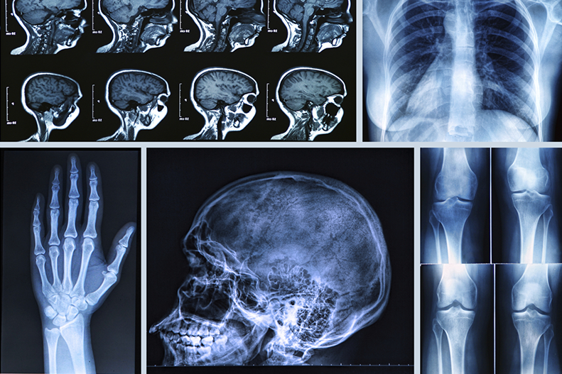
Often, patients will get a referral for a medical imaging exam. These exams can help detect diseases and identify injuries, or let doctors take a look at a certain part of the body in closer detail.
Your primary care provider can educate you about what these diagnostic imaging exams entail. Here, we discuss the differences between three commonly used imaging exams: x-rays, MRIs, and CT scans.
What Is an X-Ray?
An x-ray is an imaging test that takes pictures of your internal body structures, including bones and soft tissue.
How do x-rays work?
Doctors commonly use x-ray technology to diagnose broken bones. They can also detect pneumonia, types of cancers, and other developing conditions.
What to expect during an x-ray
In this exam, an x-ray machine sends individual x-ray particles through the body. These are recorded as digital x-ray images on a computer. These x-ray images, called “radiographs,” are a result of a shadow cast between the x-ray source and the x-ray detector to capture images of different tissues or bones inside the body.
This exam is painless. It may require you to stand still for a short period of time, which can cause temporary discomfort.
Length of x-ray exam
The time length of the x-ray depends on the body part being examined. It typically takes a matter of minutes.
What Is a Computed Tomography (CT) Procedure?
A computed tomography (CT) procedure is an imaging test that combines x-rays and computer technology to produce images of the inside of your body.
How does a CT scan work?
Doctors use CT scans primarily to look at the soft tissues of the body and various organs. A CT scan uses data from several x-ray images of structures inside the body and converts them into CT scan pictures.
CT scans can also diagnose an infection. They can also be used to guide a surgeon to the right area during a biopsy, identify masses and tumors (including cancer), and study blood vessels.
What to expect during a CT scan
During a CT scan procedure, you will lie down on a table that slides into the center of the CT scanner. Once you are inside the scanner, the machine’s x-ray beam rotates around you.
A computer creates many different CT scan images of the body, called slices. The radiologist can view these images on a monitor. You will be asked to remain still during the exam to avoid blurred images.
You may need to drink a contrast liquid for the CT scan. This allows the radiologist to highlight certain areas for a clearer image. The care team will provide the contrast will be provided to you when you arrive for your test.
Radiologists analyze the CT scan results to help diagnose disease or injury to:
- Soft tissues
- Blood vessels
- Organs
Length of CT scan
Complete scans usually take only a few minutes.
What Is Magnetic Resonance Imaging (MRI)?
A magnetic resonance imaging (MRI) exam is a diagnostic test that uses a magnetic field, radio waves, and computer technology to produce images of the inside of your body.
How does an MRI work?
MRIs detect diseases or abnormalities throughout the body, such as brain aneurysms or tumors. They also are often used as a “second look” if other imaging scans provide inconclusive results.
No radiation is used in an MRI exam. Instead, MRI images are generated by pulsing radio wave energy through the body to produce cross-sectional pictures of organs and internal structures in the body.
What to expect during an MRI
Before an MRI procedure, you will be asked to remove all metal. The purpose is to avoid interaction with the magnet inside the MRI equipment.
Some exams require a special dye (contrast), which you receive through a vein (IV) in your hand or forearm before the test. The dye helps the radiologist see certain areas more clearly.
During the scan, the technician will watch you from another room to ensure you are comfortable and give updates on the status of the exam.
Length of MRI
Depending on the area of concern, an MRI test can last between 30 and 60 minutes.
Types of MRI scans
Traditional (closed) MRI
- 23.5-inch diameter.
- May trigger claustrophobia in some patients.
- Typical exam length: 30 minutes.
Wide bore (closed) MRI
- 27.5-inch diameter.
- Better for larger patients.
- Better for claustrophobic patients.
- Typical exam length: 30 minutes.
Open MRI
- 36-inch width, 18-inch vertical.
- Ideal for larger patients.
- Ideal for claustrophobic patients.
- Typical exam length: 45 to 60 minutes.
Open upright MRI
- Nothing in front of your face, 18-inch seat.
- Ideal for larger patients.
- Ideal for claustrophobic patients.
- Typical exam length: 45 to 60 minutes.
RELATED: What Are MRI and MRA Scans?
Editor's Note: This article was originally published on , and was last reviewed on .